Retina Conditions
Macular Pucker/Epiretinal Membrane
A macular pucker is scar tissue that has formed on the eye’s macula, located in the center of the light-sensitive tissue called the retina. The macula provides the sharp, central vision we need for reading, driving, and seeing fine detail. A macular pucker can cause blurred and distorted central vision. Macular Hole
A macular hole is a small break in the macula, located in the center of the eye’s light-sensitive tissue called the retina. The macula provides the sharp, central vision we need for reading, driving, and seeing fine detail. A macular hole can cause blurred and distorted central vision. Macular holes are related to aging and usually occur in people over age 60. Vitrectomy surgery is required most of the time to repair a macular hole. Information provided courtesy of the US National Institutes of Health, National Eye Institute.
To learn more, please ask our doctors, or visit https://www.nei.nih.gov/learn-about-eye-health/eye-conditions-and-diseases |
Eye Anatomy
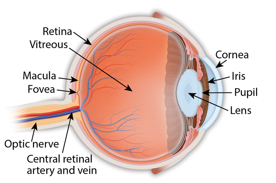
Retina: the light-sensitive membrane in the back of the eye that converts light to electrical impulses that are sent to the brain through the optic nerve
Vitreous: also called vitreous humor, a thick, colorless gel that fills the large space in the middle of the eye, behind the lens Macula: the central retina that contains the fovea Fovea: a shallow pit in the center of the retina responsible for our sharpest straight ahead vision Optic nerve: carries the message of vision from the retina to the brain Cornea: transparent convex membrane that covers the pupil and iris of the eye Iris: colored part of the eye that consists of a muscular diaphragm surrounding the pupil and regulates the light entering the eye by expanding and contracting the pupil Pupil: dark circular opening at the center of the iris in the eye, where light enters the eye Lens: focuses light to produce an image on the light-sensitive cells of the retina. Nearly spherical and convex on both sides, it sits behind the pupil |
|
Copyright © 2021 Retina Consultants. All rights reserved.
|