Retina Conditions
Retinal Detachment
Retinal detachment is an eye problem that happens when your retina (a light-sensitive layer of tissue in the back of your eye) is pulled away from its normal position at the back of your eye. In some cases there may be small areas of the retina that are torn. These areas, called retinal tears or retinal breaks, can lead to retinal detachment. What are the symptoms of retinal detachment? Not all patients with retinal detachment have the same symptoms. In fact, some patients have no warning signs at all. These are the warning signs: First, a sudden increase in the number or size of floaters in the vision is common. This may be accompanied by awareness of light flashes in the side vision. Finally, the sensation of a curtain or veil over part of the vision is usually alarming enough to get most people to call for an exam. It is important to recognize a retinal tear or detachment as promptly as possible because the problem may be easier to correct if caught in its early stages. If scleral buckling surgery, vitrectomy or a combination of vitrectomy with scleral buckle is required then surgery would be scheduled in a timely fashion. Information provided courtesy of the US National Institutes of Health, National Eye Institute.
To learn more, please ask our doctors, or visit https://www.nei.nih.gov/learn-about-eye-health/eye-conditions-and-diseases |
Eye Anatomy
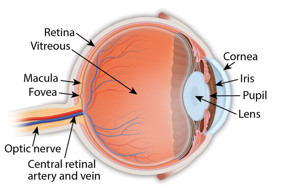
Retina: the light-sensitive membrane in the back of the eye that converts light to electrical impulses that are sent to the brain through the optic nerve
Vitreous: also called vitreous humor, a thick, colorless gel that fills the large space in the middle of the eye, behind the lens Macula: the central retina that contains the fovea Fovea: a shallow pit in the center of the retina responsible for our sharpest straight ahead vision Optic nerve: carries the message of vision from the retina to the brain Cornea: transparent convex membrane that covers the pupil and iris of the eye Iris: colored part of the eye that consists of a muscular diaphragm surrounding the pupil and regulates the light entering the eye by expanding and contracting the pupil Pupil: dark circular opening at the center of the iris in the eye, where light enters the eye Lens: focuses light to produce an image on the light-sensitive cells of the retina. Nearly spherical and convex on both sides, it sits behind the pupil |
|
Copyright © 2021 Retina Consultants. All rights reserved.
|