RETINA CONSULTANTS

401-274-5844

  • Home
  • For Patients
    • Office Visits
    • Office Testing
    • Surgical Patients
    • Patient Forms
  • Our Practice
    • About Us
    • Office Policies
    • Payment
    • Contact Us
  • Conditions
    • AMD: Age-related Macular Degeneration
    • Diabetic Retinopathy
    • Retinal Detachment
    • Retinal Vein Occlusions
    • Macular Pucker and Macular Hole
    • CSR: Central Serous Retinopathy
    • Vitreous Detachment
    • Floaters
  • Treatments
    • Intravitreal Injections
    • Laser Photocoagulation
    • Cryotherapy/Cryopexy
    • PDT: Photodynamic Therapy
    • Surgical Procedures
  • Videos
    • Dilating Drops, Angiography
    • Diabetic Eye Disease
    • Detached Retina and Torn Retina
    • Age-related Macular Degeneration (AMD)
    • Other Macular Conditions
    • Vein Occulsions
  • Patient Portal
  • COVID-19

Retina Conditions

Vitreous Detachment
The vitreous is the gel-like fluid that fills your eye. It’s full of tiny fibers that attach to your retina (the light-sensitive layer of tissue at the back of the eye). As you get older, the fibers of your vitreous pull away from the retina. This is called vitreous detachment. It usually happens after age 50. If you notice symptoms that affect your vision, talk to your eye doctor.

Information provided courtesy of the US National Institutes of Health, National Eye Institute.

To learn more, please ask our doctors, or visit
https://www.nei.nih.gov/learn-about-eye-health/eye-conditions-and-diseases

Eye Anatomy

Picture
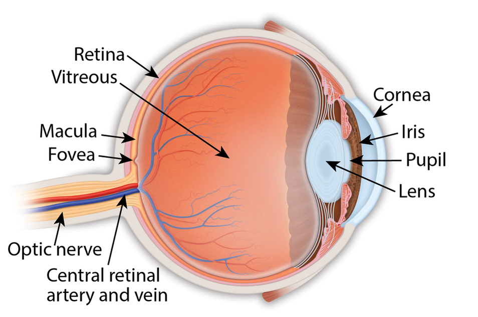
Retina: the light-sensitive membrane in the back of the eye that converts light to electrical impulses that are sent to the brain through the optic nerve

Vitreous: also called vitreous humor, a thick, colorless gel that fills the large space in the middle of the eye, behind the lens

Macula: the central retina that contains the fovea

Fovea: a shallow pit in the center of the retina responsible for our sharpest straight ahead vision

Optic nerve: carries the message of vision from the retina to the brain

Cornea: transparent convex membrane that covers the pupil and iris of the eye

Iris: colored part of the eye that consists of a muscular diaphragm surrounding the pupil and regulates the light entering the eye by expanding and contracting the pupil

Pupil: dark circular opening at the center of the iris in the eye, where light enters the eye

Lens: focuses light to produce an image on the light-sensitive cells of the retina. Nearly spherical and convex on both sides, it sits behind the pupil
Legal Disclaimer
Copyright © 2021 Retina Consultants. All rights reserved.
  • Home
  • For Patients
    • Office Visits
    • Office Testing
    • Surgical Patients
    • Patient Forms
  • Our Practice
    • About Us
    • Office Policies
    • Payment
    • Contact Us
  • Conditions
    • AMD: Age-related Macular Degeneration
    • Diabetic Retinopathy
    • Retinal Detachment
    • Retinal Vein Occlusions
    • Macular Pucker and Macular Hole
    • CSR: Central Serous Retinopathy
    • Vitreous Detachment
    • Floaters
  • Treatments
    • Intravitreal Injections
    • Laser Photocoagulation
    • Cryotherapy/Cryopexy
    • PDT: Photodynamic Therapy
    • Surgical Procedures
  • Videos
    • Dilating Drops, Angiography
    • Diabetic Eye Disease
    • Detached Retina and Torn Retina
    • Age-related Macular Degeneration (AMD)
    • Other Macular Conditions
    • Vein Occulsions
  • Patient Portal
  • COVID-19