Retina Conditions
Retinal Vein Occlusions
Retinal vein occlusion is a blockage of the veins in the retina. There are two forms of retinal vein occlusion, both require treatment to prevent vision loss and other complications. Central retinal vein occlusion (CRVO) occurs when the main retinal vein is blocked, and may result in bleeding and swelling of the retina or macula, leading to a severe decrease in vision. Abnormal vessels may develop, and if they grow on the iris, a severe and a painful increase in eye pressure may develop. Branch retinal vein occlusion (BRVO) occurs when a branch of the main retinal vein becomes blocked, reducing blood flow to a portion of the retina, causing vision loss, although usually not as severe as in CRVO. Macular edema may develop with a BRVO as well. Retinal vein occlusion may be treated with intraocular injections and laser. Information provided courtesy of the US National Institutes of Health, National Eye Institute.
To learn more, please ask our doctors, or visit https://www.nei.nih.gov/learn-about-eye-health/eye-conditions-and-diseases |
Eye Anatomy
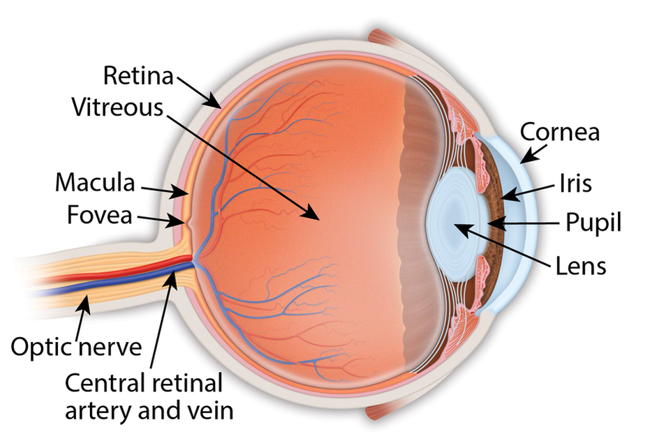
Retina: the light-sensitive membrane in the back of the eye that converts light to electrical impulses that are sent to the brain through the optic nerve
Vitreous: also called vitreous humor, a thick, colorless gel that fills the large space in the middle of the eye, behind the lens Macula: the central retina that contains the fovea Fovea: a shallow pit in the center of the retina responsible for our sharpest straight ahead vision Optic nerve: carries the message of vision from the retina to the brain Cornea: transparent convex membrane that covers the pupil and iris of the eye Iris: colored part of the eye that consists of a muscular diaphragm surrounding the pupil and regulates the light entering the eye by expanding and contracting the pupil Pupil: dark circular opening at the center of the iris in the eye, where light enters the eye Lens: focuses light to produce an image on the light-sensitive cells of the retina. Nearly spherical and convex on both sides, it sits behind the pupil |
|
Copyright © 2021 Retina Consultants. All rights reserved.
|